
Congenital ear canal atresia with extensive ceruminolith
Name: Winter | Congenital ear canal atresia with extensive ceruminolith formation and secondary Malassezia otitis externa due to impaired self-cleaning mechanisms.
Name: Winter Gelle
Age: 1 year old
Breed: Australian Shepherd
History: Winter presented for evaluation of aural pruritus. On examination, the opening of the external ear canal was compromised and a regular otoscopic examination could not be performed.
Test /Procedure: A CT scan revealed a narrow membrane at the opening of the left external auditory canal with extensive obstructive debris in the external ear canal extending to the tympanic membrane. There were no structural abnormalities.
Diagnosis: Congenital ear canal atresia with extensive ceruminolith formation and secondary Malassezia otitis externa due to impaired self-cleaning mechanisms.
Treatment: The membrane was surgically excised to permit oto-endoscopic evaluation and irrigation of the external auditory canal. A large amount of compacted, brown debris was manually removed and the external ear canal irrigated. A ceruminolytic was used to aid breakdown of the remaining debris adherent to the macerated canal wall. Cytology of the debris revealed a significant amount of Malassezia.
The ear canal and tympanic membrane have a self-cleaning and repair mechanism referred to as epithelial migration, whereby the epithelial cells adjacent to the tympanic membrane migrate distally to remove excess cerumen and exudate out of the ear canal. In this case, the ear canal atresia has impaired the extrusion of normal cerumen, leading to accumulation, compaction and maceration of the canal walls, creating a warm and humid environment – a perfect breeding ground for Malassezia.
Prevention or Follow up: Following the excision of the membrane, normal self-cleaning mechanisms are expected to recover. If regrowth of the membrane occurs, a corrective laser procedure may be warranted.

Image 1: Winter displaying low left ear carriage

Image 2: CT scan of Winter’s head demonstrating a thin soft tissue membrane bridging across the opening of the external ear canal (red arrow) and soft tissue density within the external ear canal (white arrow)

Image 3: Thin membrane forming a narrowed entrance to the external ear canal (left ear)
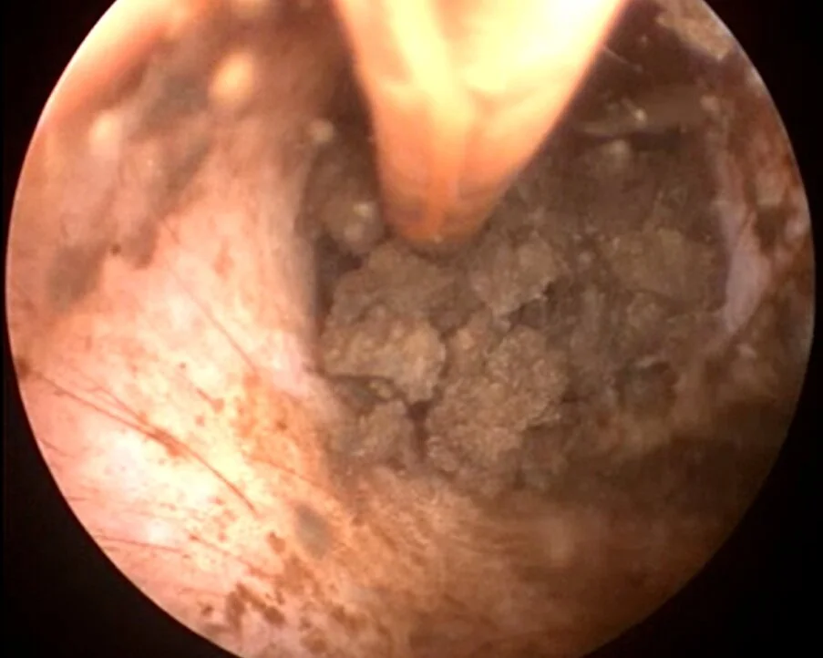
Image 4: Extensive ceruminolith formation being flushed out during oto-endoscopic ear flush

Image 5: Cytology of Winter's left ear revealing overgrowth of Malassezia